Implant supported fixed hybrids, also known as “bone anchored” bridges have been a well-studied treatment approach for the rehabilitation of the completely edentulous patient for over 30 years.1 Research on the technique began in Sweden in the 1950’s by Brånemark and colleagues, and the phenomenon of “osseointegration” was eventually introduced to North America in 1982.2 The original protocol proposed by Brånemark was necessarily rigid and precise yielding favorable implant and prosthesis survival rates. The technique as originally introduced, called for the placement of six implants in the completely edentulous jaw between the sinuses in the maxilla, or mental foramens in the mandible to support a cast framework enforced denture with bilateral distal extension cantilevers. Since that time the concept of osseointegration has evolved and has been applied to the partially edentulous patient including single tooth replacement with exceptional success. The study of dental implants and associated prosthetics is one of the most highly researched areas in all of dentistry and one of the most significant advances in dental patient management of the 20th century.
Recent studies now provide adequate evidence supporting the use of as few as 4 implants to retain complete fixed hybrid prostheses for both the maxilla and mandible.3-4 The technique, also referred to as “All on Four” involves the placement of 4 or more implants between the sinuses in the maxilla, or mental foramens in the mandible, with the most distal implants angled in such a way as to reduce the cantilever length of the final prosthesis. The implants are immediately loaded with a provisional prosthesis shortly after placement, and usually on the same day. The benefit of this technique includes the use of fewer implants, avoidance of grafting, less invasiveness, less treatment time, less cost, and immediate function. With immediate function, a provisional conventional denture is also avoided.
The technique is indicated for patients that are completely edentulous, or have a terminal dentition in one or both jaws. It is also indicated when salvaging teeth is financially impractical or undesirable, such as when extensive or invasive procedures are required to retain them, or when retaining compromised teeth will result in an unfavorable prosthetic prognosis (Fig. 1)
 Figure 1 – Rampant tooth decay as a result of severe xerostomia. Retaining the salvageable teeth would lead to a compromised prosthetic prognosis due to the chronic xerostomia as well as increase treatment time and cost.
 Figure 2 – Pre surgical planning includes determination of inner-occlusal space requirements, need for lip support and amount of bone resection (outlined in red) as well as implant size, position and angulation.
Figure 3 – Multiple surgical templates including the conversion prosthesis are fabricated prior to surgery.
An accurate diagnosis and a thorough pre-treatment plan are essential for success. Prior to surgery, diagnostic study models are made and mounted on a semi-adjustable articulator using a face bow transfer. Diagnostic wax-ups are created and the prosthetic plan is finalized. Using CBCT and other imaging, the surgical procedures are then planned, with careful attention given to the amount of inner-occlusal space required for all the surgical and prosthetic components as well as the desired location of the transition line between the prosthesis and oral tissue (Fig. 2). For proper esthetics, the transition line must be hidden beyond the high smile lip position. In addition, the need for lip support and a denture flange is evaluated. If lip support is required, an overdenture rather than a fixed prosthesis is usually indicated. Based on the diagnostic wax-ups and pretreatment planning, multiple surgical templates are then fabricated including a bone reduction template, an implant surgical guide as well as a conversion prosthesis (Fig. 3).

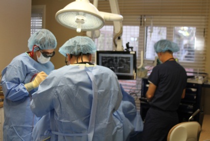
Figure 4 – Surgery is performed in a properly equipped suite.

Figure 5 – Surgery involves a sterile technique under moderate sedation.
Surgery is performed in a properly equipped surgical suite (Fig. 4) using a sterile technique, typically under moderate sedation (Fig. 5). The surgical procedure typically takes approximately 1-2 hours per jaw depending on how many teeth are removed, the extent of alveolectomy and the number of implants placed. Following implant placement, prosthetic abutments are attached; tissues are closed, and implant transfer impressions are made by the surgeon (Fig. 6). Once the patient has recovered from the anesthesia, they are transported to the office of the prosthodontist or restorative dentist for the immediate load conversion prosthesis delivery.

Figure 6 – Implant placement and abutment connection following an “All on Four” approach in the maxilla.
At the office of the restorative dentist, transfer impressions are poured and master casts of the implants and tissues are made to facilitate fabrication of the conversion prostheses. Provisional copings are then attached to the implants intra-orally. The conversion prosthesis is carefully adjusted to ensure proper seating over the provisional copings at the pre-determined vertical dimension of occlusion.The provisional copings are then luted to the conversion prosthesis intra-orally, and occlusal registrations are made. The conversion prosthesis with the luted provisional copings is then taken back to the laboratory for final finish and polish and prepared for insertion and immediate loading (Fig. 7).

Figure 7 – The conversion prosthesis is luted to the provisional abutments intra-orally, finished and polished and prepared for immediate loading.
Following appropriate healing time for implant integration, conventional implant prosthetic techniques are used to fabricate the final fixed hybrid prostheses (Figs. 8 and 9). Fixed hybrid bridges can be made with cast frameworks, or CAD-CAM milled titanium or zirconia frameworks. Hybrid bridges have historically been constructed using bonded pink acrylic resin and denture teeth. However, emerging laboratory techniques can also provide monolithic zirconia bridges without metal frameworks.

Figure 8 – Final prostheses delivery with an “All on Four” hybrid in the maxilla and a lower hybrid supported by five implants.

Figure 9 – A patient with an “All on Four” hybrid in the mandible opposing a complete maxillary denture.
A recent retrospective evaluation of 800 implants placed in 200 jaws of 152 patients treated with the “All on Four” technique reported a 96% implant survival rate in the maxilla and a 98% implant survival rate in the mandible. An identical survival rate for the tilted vs. non tilted implants was found over a 6 year evaluation period. The authors conclude that the “All on Four” approach is a viable treatment option for indicated patients.5 The overall technique is complex however and not recommended for untrained or inexperienced practitioners.
References
- Branemark PI, Zarb GA, Albrektsson T. Tissue-integrated prostheses: Osseointegration in clinical dentistry. Hanover Park, IL: Quintessence Publishing; 1985
- Adell R, Lekholm U, Rockler B, Branemark PI. A 15-year study of osseointegrated implants in the treatment of the edentulous jaw. Int J Oral Surg 1981;10(6):387-416
- Malo P, de Araujo Nobre M, Lopes A, Moss SM, Molina GJ 2011; A longitudinal study of the survival of All-on-4 implants in the mandible with up to 10 years of follow-up. JADA 142(3):310-320.
- Malo P, de Araujo Nobre M, Lopes A, Francischone C, Rigolizzo M. “All-on-4” immediate-function concept for completely edentulous maxillae: a clinical report on the medium (3 years) and long-term (5 years) outcomes. Clin Implant Dent Relat Res 2012; 14 (suppl 1):e139-150.
- Balshi TJ, Wolfinger GJ, Slauch RW, Balshi SF. A retrospective analysis of 800 Brånemark system implants following the All on Four protocol. J of Prosthodontics 2014; 23:83-88.

